Foot Muscles Mri / Mri Of The Muscles In Wohlfart Kugelberg Welander Disease Journal Of The Neurological Sciences
Hi, i had surgery on my shoulder about 8 years ago and have two metal anchors in my shoulder. Indications for foot mri scan. ► hip ► pelvis ► thigh ► knee ► lower extremity/shin ► ankle ► foot. Mri with hardware in foot? Bone contusions, osteonecrosis, marrow oedema syndromes, and stress > fractures) > synovial based disorders ( e.g.
Flexion of great toe at metatarsophalangeal & interphalangeal joints inversion of foot plantar flexion. Muscles of the foot are located on its rear and on the sole. Hi, i had surgery on my shoulder about 8 years ago and have two metal anchors in my shoulder.
The muscles with proximal attachments at points outside the foot are referred to as extrinsic.
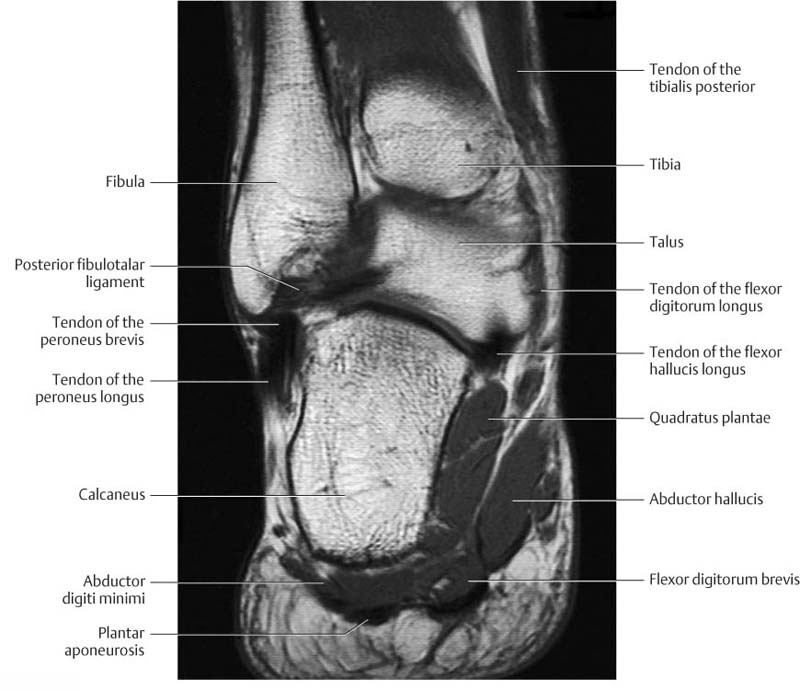
Thank you for your attention. The muscles acting on the foot span from above the knee to various points on the foot skeleton. Mri with hardware in foot? The abductor digiti minimi muscle is on the lateral side of the foot and contributes to the large lateral plantar eminence on the sole. Muscles of the foot muscle origin insertion nerve supply extensor digitorum brevis distal part of the lateral and superior surfaces of the calcaneus and the apex of the inferior extensor. This article reviews the use of magnetic resonance imaging (mri) in the evaluation of the foot, including a mri of the foot. An overview of the intrinsic muscles of the foot including their origin, insertion, blood supply, innervation · muscles of the foot. Subscribe to foot & ankle problems. Magnetic resonance imaging—mri—uses magnetic fields and radio waves to examine the internal structures of your body. Gray's anatomy for students, 2nd ed. Learn about foot and ankle mri here. Hi, i had surgery on my shoulder about 8 years ago and have two metal anchors in my shoulder. If you'd like to support us and get something great in return. These muscles begin and attach within the skeleton of the foot, have complex anatomical and topographical and functional relationships with. A magnetic resonance imaging (mri) was performed on a normal subject;
The muscles acting on the foot span from above the knee to various points on the foot skeleton. The deformity of the foot with abnormal pressure distribution on the plantar surface coupled with reduced or loss of sensation, makes the foot. Intrinsic muscles of the feet part 2 | layers 3 & 4. Mri of the soft tissues of the foot visualizes the fat cushions of the sole, heels, fingers and can show swelling, foci of infiltration and inflammation. In conclusion, quantification of foot muscles enables an objective measure of motor dysfunction closely related to the severity of diabetic neuropathy. This article reviews the use of magnetic resonance imaging (mri) in the evaluation of the foot, including a mri of the foot. What to look for, where to look & how to report.
Intrinsic muscles of the feet part 2 | layers 3 & 4.
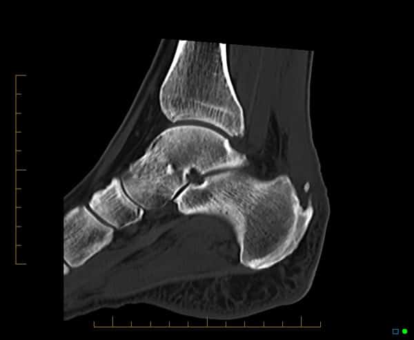
The extrinsic muscles are located in the anterior and lateral compartments of the leg. If you'd like to support us and get something great in return. Gray's anatomy for students, 2nd ed. 12 photos of the foot muscle anatomy mri. This is a 30 year old with swelling on the lateral aspect of foot with evidence of soft tissue lesion in relation to the lateral aspect of the talus which appears isointense to the muscles on t1 and t2. ► shoulder ► elbow ► wrist ► finger ► thumb. Mri with hardware in foot? Muscles of the foot are located on its rear and on the sole. Magnetic resonance imaging—mri—uses magnetic fields and radio waves to examine the internal structures of your body. Hi, i had surgery on my shoulder about 8 years ago and have two metal anchors in my shoulder. Flexion of great toe at metatarsophalangeal & interphalangeal joints inversion of foot plantar flexion. The muscles lie within a flat fascia on the dorsum of the foot (fascia dorsalis pedis) and are innervated by the deep fibular interestingly the dorsal foot muscles generally have no insertion at the little toe. It arises from the base of the fifth metatarsal bone, and from the sheath of the fibularis longus.
A magnetic resonance imaging (mri) was performed on a normal subject; This article reviews the use of magnetic resonance imaging (mri) in the evaluation of the foot, including a mri of the foot. Posted by radiologyer at 8:12 am. Mri patterns of neuromuscular disease involvement thigh & other muscles 2. Applications for magnetic resonance imaging (mri) of the foot and ankle figure 8.4 image planes for foot and ankle mri.
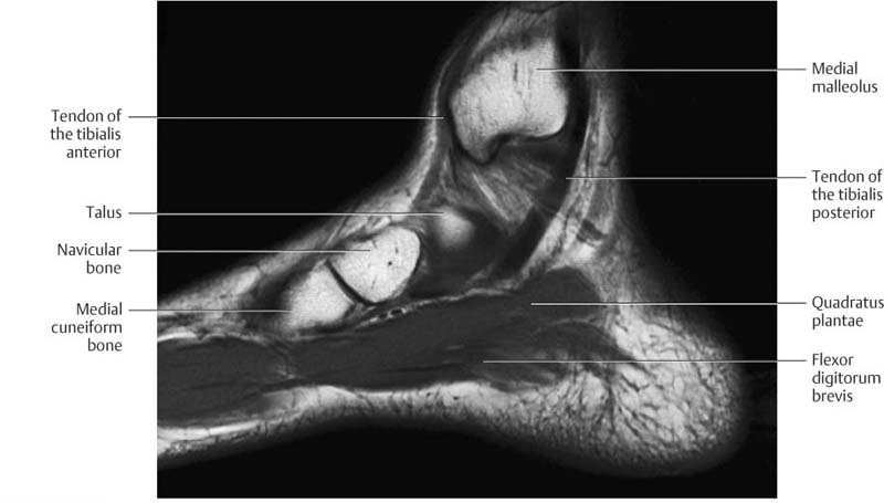
Muscles of the foot muscle origin insertion nerve supply extensor digitorum brevis distal part of the lateral and superior surfaces of the calcaneus and the apex of the inferior extensor.
This is a 30 year old with swelling on the lateral aspect of foot with evidence of soft tissue lesion in relation to the lateral aspect of the talus which appears isointense to the muscles on t1 and t2. Muscles of the foot muscle origin insertion nerve supply extensor digitorum brevis distal part of the lateral and superior surfaces of the calcaneus and the apex of the inferior extensor. Learn about foot and ankle mri here. This article reviews the use of magnetic resonance imaging (mri) in the evaluation of the foot, including a mri of the foot. Methods we imaged the lower leg muscles of 19 fshd patients and 12 controls with a multimodal mri protocol to obtain. Mri with hardware in foot? Posted by radiologyer at 8:12 am. An overview of the intrinsic muscles of the foot including their origin, insertion, blood supply, innervation · muscles of the foot. Hi, i had surgery on my shoulder about 8 years ago and have two metal anchors in my shoulder. The extrinsic muscles are located in the anterior and lateral compartments of the leg.

Upper and lower lines mark.
 was performed on a normal subject;")
It arises from the base of the fifth metatarsal bone, and from the sheath of the fibularis longus.

Foot positioned for axial images of the ankles;

Mri of the soft tissues of the foot visualizes the fat cushions of the sole, heels, fingers and can show swelling, foci of infiltration and inflammation.

Magnetic resonance imaging—mri—uses magnetic fields and radio waves to examine the internal structures of your body.

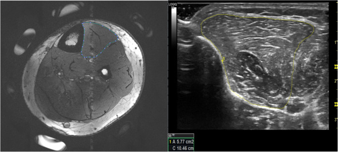
In conclusion, quantification of foot muscles enables an objective measure of motor dysfunction closely related to the severity of diabetic neuropathy.

Bone contusions, osteonecrosis, marrow oedema syndromes, and stress > fractures) > synovial based disorders ( e.g.

New msk mri course online foot & toes 2021 covers the complex anatomy & common abnormalities in reporting.

Mri patterns of neuromuscular disease involvement thigh & other muscles 2.

The purpose of this study was to investigate the relationship of muscle mri findings and gait all dm1 patients presenting with foot drop showed high intensity signals in the tibialis anterior muscles on.

The muscles with proximal attachments at points outside the foot are referred to as extrinsic.

Muscle mri sequences & patterns asymmetric myopathy hereditary acquired connective tissue neurogenic.
► shoulder ► elbow ► wrist ► finger ► thumb.

Muscles of the foot are located on its rear and on the sole.

Muscles of the foot are located on its rear and on the sole.

Muscles of the ankle and foot.

By muhammad ali, mb bs;

Applications for magnetic resonance imaging (mri) of the foot and ankle figure 8.4 image planes for foot and ankle mri.

By muhammad ali, mb bs;

Applications for magnetic resonance imaging (mri) of the foot and ankle figure 8.4 image planes for foot and ankle mri.

What to look for, where to look & how to report.

By muhammad ali, mb bs;

The muscles acting on the foot can be divided into two distinct groups;

A magnetic resonance imaging (mri) was performed on a normal subject;
 tests involve taking images of the foot the mri machine uses radio wave energy pulses and a magnetic field to produce the foot and ankle images.")
Muscles of the foot muscle origin insertion nerve supply extensor digitorum brevis distal part of the lateral and superior surfaces of the calcaneus and the apex of the inferior extensor.

Thank you for your attention.

Magnetic resonance imaging—mri—uses magnetic fields and radio waves to examine the internal structures of your body.

Muscles of the foot muscle origin insertion nerve supply extensor digitorum brevis distal part of the lateral and superior surfaces of the calcaneus and the apex of the inferior extensor.

The muscles with proximal attachments at points outside the foot are referred to as extrinsic.
 and are innervated by the deep fibular interestingly the dorsal foot muscles generally have no insertion at the little toe.")
Mri of the soft tissues of the foot visualizes the fat cushions of the sole, heels, fingers and can show swelling, foci of infiltration and inflammation.

Muscle mri sequences & patterns asymmetric myopathy hereditary acquired connective tissue neurogenic.

Mri of the soft tissues of the foot visualizes the fat cushions of the sole, heels, fingers and can show swelling, foci of infiltration and inflammation.

Posting Komentar untuk "Foot Muscles Mri / Mri Of The Muscles In Wohlfart Kugelberg Welander Disease Journal Of The Neurological Sciences"